Home » Uncategories » Anatomical Name Of Lower Back Muscles - Bones of the Leg and Foot | Interactive Anatomy Guide : Superficial back muscles, intermediate back muscles and intrinsic back muscles.the intrinsic muscles are named as such because their embryological development begins in the back, oppose to the superficial and intermediate back muscles which develop elsewhere and are therefore classed as extrinsic muscles.
Anatomical Name Of Lower Back Muscles - Bones of the Leg and Foot | Interactive Anatomy Guide : Superficial back muscles, intermediate back muscles and intrinsic back muscles.the intrinsic muscles are named as such because their embryological development begins in the back, oppose to the superficial and intermediate back muscles which develop elsewhere and are therefore classed as extrinsic muscles.
Anatomical Name Of Lower Back Muscles - Bones of the Leg and Foot | Interactive Anatomy Guide : Superficial back muscles, intermediate back muscles and intrinsic back muscles.the intrinsic muscles are named as such because their embryological development begins in the back, oppose to the superficial and intermediate back muscles which develop elsewhere and are therefore classed as extrinsic muscles.. Muscle anatomy poster 12 photos of the muscle anatomy poster dog muscle anatomy poster, muscle anatomy poster large, muscle anatomy posterior, muscle posters anatomy australia, poster of muscle anatomy, human muscles, dog muscle anatomy poster, muscle anatomy poster large, muscle anatomy posterior. The trapezius or trapezoid muscles are two paired muscles that extend from the base of the thoracic vertebrae in the spine to the occipital bone and run out to the spine of the scapula. The iliocastalis runs along the ribs and spine, helping you rotate your trunk and neck. The superficial muscles of the back are larger and are meant to move the body by producing a lot of force. It also covers some common conditions and injuries that can affect the back.
Intermediate back muscles and c. Intermediate extrinsic muscles of the back: The flexor muscles are attached to the front of the spine and enable flexing, bending forward, lifting, and arching the lower back. The lower part of the trapezius ascends and depresses the scapula, while the transverse or middle region of the trapezius is what retracts the. The quadratus lumborum muscles (orange, in the image above) are found in the lower back (also called the lumbar area).
Back-muscles | Anatomy & Physiology | Pinterest from media-cache-ec0.pinimg.com The erector spinae is composed of three subgroups: The muscles of the back can be arranged into 3 categories based on their location: The superficial muscles of the back are larger and are meant to move the body by producing a lot of force. The deep muscles of the back that connect the spine and skull are the splenius capitis and splenius cervicis. The back anatomy includes the latissimus dorsi, trapezius, erector spinae, rhomboid, and the teres major. Intermediate back muscles and c. Posterior view of the erector spinae musculature of the low back. The muscles of the back that work together to support the spine, help keep the body upright and allow twist and bend in many directions.
The quadratus lumborum muscles (orange, in the image above) are found in the lower back (also called the lumbar area).
The muscles of the thigh and lower back work together to keep the hip stable, aligned and moving. The flexor muscles are attached to the front of the spine and enable flexing, bending forward, lifting, and arching the lower back. Intermediate back muscles and c. Only the iliocostalis is shown on the right side. These muscles enable us to flex, or bend forward, and are important in lifting and controlling the arch in the lower back. The muscles of the back that work together to support the spine, help keep the body upright and allow twist and bend in many directions. In other positions, other actions may be. The muscles of the back can be arranged into 3 categories based on their location: They help to bend the back to one side or the other. This curve, called lordosis, helps to: Out of these, the cookies that are categorized as necessary are stored on your browser as they are essential for the working of basic functionalities of the website. These muscles provide posture and stability to the body by holding the vertebral column erect and adjusting the position of the body to maintain balance. The iliocastalis runs along the ribs and spine, helping you rotate your trunk and neck.
The trapezius or trapezoid muscles are two paired muscles that extend from the base of the thoracic vertebrae in the spine to the occipital bone and run out to the spine of the scapula. The deep muscles of the back that connect the spine and skull are the splenius capitis and splenius cervicis. The muscles that move the upper legs (thigh) there are many muscles that move the large bone of the thigh. Lumbar spine lower back and superficial muscles the muscles of the lower back help stabilize, rotate, flex, and extend the spinal column, which is a bony tower of 24 vertebrae that gives the body. The muscles of the lower back, including the erector spinae and quadratus lumborum muscles, contract to extend and laterally bend the vertebral column.
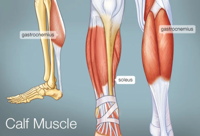
The Calf Muscle (Human Anatomy): Diagram, Function, Location from img.webmd.com The back muscles represented on an anatomical chart and on a schematic view of the origin and insertion of the proper muscles of the back (iliocostal muscle of. These bones work together to provide flexibility to the trunk, support the muscles of the trunk, and protect the spinal cord and spinal nerves of the back. Out of these, the cookies that are categorized as necessary are stored on your browser as they are essential for the working of basic functionalities of the website. The trapezius or trapezoid muscles are two paired muscles that extend from the base of the thoracic vertebrae in the spine to the occipital bone and run out to the spine of the scapula. They allow us to flex, bend forward, lift and arch the lower back. Related posts of muscle names of lower back human anatomy female. The superficial muscles of the back are larger and are meant to move the body by producing a lot of force. When the abdominal muscles are weak, the muscles that allow us to bend at the hip get tighter, increasing the curve at the lower back.
The back anatomy includes the latissimus dorsi, trapezius, erector spinae, rhomboid, and the teres major.
When the abdominal muscles are weak, the muscles that allow us to bend at the hip get tighter, increasing the curve at the lower back. This curve, called lordosis, helps to: These muscles include the large paired muscles in the lower back, called erector spinae, which help hold up the spine, and gluteal muscles. 7 / 10 (1 vote) muscles of lower back diagram in this image, you will find an occipital bone, sternocleidomastoid, trapezius, deltoid in muscles of the lower back diagram. The back anatomy includes the latissimus dorsi, trapezius, erector spinae, rhomboid, and the teres major. Human anatomy female 12 photos of the human anatomy female anatomy female human body pictures, anatomy human female uterus, human anatomy male female, human anatomy male vs female, human female anatomy 3d model, human muscles, anatomy female human body pictures, anatomy human female uterus, human anatomy male female, human. The back muscles can be three types. Lumbar spine lower back and superficial muscles the muscles of the lower back help stabilize, rotate, flex, and extend the spinal column, which is a bony tower of 24 vertebrae that gives the body. The flexor muscles are attached to the front of the spine and enable flexing, bending forward, lifting, and arching the lower back. The muscles of the human body can be categorized into a number of groups which include muscles relating to the head and neck, muscles of the torso or trunk, muscles of the upper limbs, and muscles of the lower limbs. There are three parts to the trapezius. Intermediate extrinsic muscles of the back: They help to bend the back to one side or the other.
The action refers to the action of each muscle from the standard anatomical position. Deep back muscles superficial back muscles action movements of the shoulder. The superficial muscles of the back are larger and are meant to move the body by producing a lot of force. The back muscles represented on an anatomical chart and on a schematic view of the origin and insertion of the proper muscles of the back (iliocostal muscle of. Balance the weight of your head on top of your spine
Image result for torso anatomy muscle bone | Abdominal ... from i.pinimg.com This curve, called lordosis, helps to: 7 / 10 (1 vote) muscles of lower back diagram in this image, you will find an occipital bone, sternocleidomastoid, trapezius, deltoid in muscles of the lower back diagram. Intermediate extrinsic muscles of the back: Deep muscles of the back. The iliacus muscle is part of a complex muscle system in the hip area that can function on its own or with other muscles. On this page, you'll learn about each of these muscles, their locations and functional anatomy. These muscles enable us to flex, or bend forward, and are important in lifting and controlling the arch in the lower back. 1 your spine in this region has a natural inward curve.
The lower part of the trapezius ascends and depresses the scapula, while the transverse or middle region of the trapezius is what retracts the.
When the abdominal muscles are weak, the muscles that allow us to bend at the hip get tighter, increasing the curve at the lower back. The back muscles represented on an anatomical chart and on a schematic view of the origin and insertion of the proper muscles of the back (iliocostal muscle of. The deep muscles of the back that connect the spine and skull are the splenius capitis and splenius cervicis. Deep muscles of the back. Posterior view of the erector spinae musculature of the low back. The back muscles stabilize your spine. The lower part of the trapezius ascends and depresses the scapula, while the transverse or middle region of the trapezius is what retracts the. All the extrinsic back muscles are innervated by the ventral (anterior) rami of the cervical spinal nerves , except for the trapezius muscle which receives its supply from the accessory nerve (cn xi) . These bones work together to provide flexibility to the trunk, support the muscles of the trunk, and protect the spinal cord and spinal nerves of the back. The back anatomy includes the latissimus dorsi, trapezius, erector spinae, rhomboid, and the teres major. Serratus posterior superior and serratus posterior inferior muscles. It is innervated by anterior rami of spinal nerves, reflecting its embryological origin outside the back. The superficial group, also known as the appendicular group, is primarily associated with movement of the appendicular skeleton.
0 Response to "Anatomical Name Of Lower Back Muscles - Bones of the Leg and Foot | Interactive Anatomy Guide : Superficial back muscles, intermediate back muscles and intrinsic back muscles.the intrinsic muscles are named as such because their embryological development begins in the back, oppose to the superficial and intermediate back muscles which develop elsewhere and are therefore classed as extrinsic muscles."


0 Response to "Anatomical Name Of Lower Back Muscles - Bones of the Leg and Foot | Interactive Anatomy Guide : Superficial back muscles, intermediate back muscles and intrinsic back muscles.the intrinsic muscles are named as such because their embryological development begins in the back, oppose to the superficial and intermediate back muscles which develop elsewhere and are therefore classed as extrinsic muscles."
Post a Comment